In a longitudinal research printed in Scientific Experiences, researchers examine the impact of maternal bodily exercise on the scale and progress of the yolk sac throughout early being pregnant. To this finish, maternal bodily exercise was discovered to have an effect on human yolk sac improvement, with this impact depending on gestational age and embryonic intercourse.
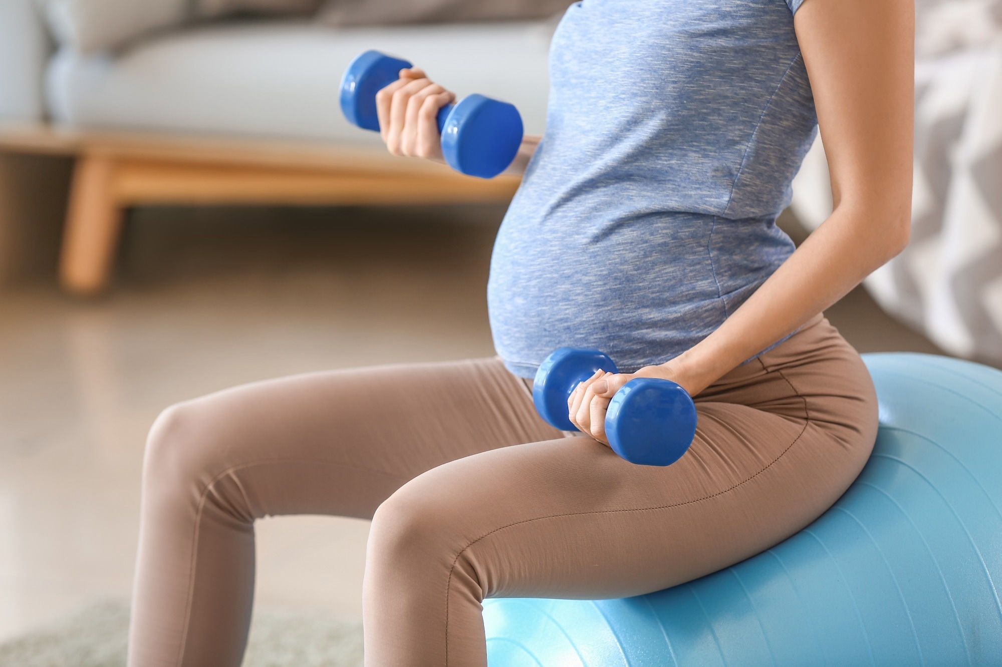
 Examine: Maternal bodily exercise impacts yolk sac dimension and progress in early being pregnant, however ladies and boys use totally different methods. Picture Credit score: Pixel-Shot / Shutterstock.com
Background
The yolk sac is a construction noticed in the course of the early improvement of the human embryo that gives vitamins and permits fuel alternate to the fetus till the placenta develops totally. Furthermore, the yolk sac additionally performs a job in total developmental processes akin to protein synthesis, gastrointestinal tract formation, stem cell manufacturing, and hematopoiesis.
Varied maternal components akin to peak, weight achieve, and sleep period are recognized to have an effect on yolk sac dimension. Maternal bodily exercise, for instance, is a modifiable life-style issue related to glucose management, weight achieve, and total outcomes for the mom and the fetus.
In regards to the research
The current research aimed to grasp how maternal bodily exercise probably impacts the intrauterine setting and early embryonic improvement, as indicated by the scale and progress of the yolk sac.
As part of the continuing conception-implantation interval in being pregnant (CONIMPREG) analysis program, this potential and longitudinal research included 196 wholesome and nonsmoking girls with common menstrual cycles who might conceive naturally. The ladies had been between 20 and 35 years outdated, and their physique mass index (BMI) was between 18 and 30 kg/m2.
The evaluation of members was performed at 4 research visits. Through the first go to, previous to conception, peak and physique composition had been measured, and bodily exercise was recorded utilizing an actigraph, a wi-fi and noninvasive monitor.
Within the second go to at seven weeks of gestation, embryo viability, gestation size, and yolk sac had been assessed by means of ultrasound imaging. At 10 weeks, yolk sac measures had been repeated. Through the closing go to at 13 weeks, maternal bodily exercise and physique composition had been reevaluated.
The statistical evaluation concerned utilizing strange least sq. linear (OLS) regression fashions, evaluation of variance (ANOVA), and the estimation of means, customary deviations, minima, and maxima.
Examine findings
The imply gestational size was 281 days in line with the final menstrual interval (LMP) date or 278.5 days, as mirrored by the crown-rump size (CRL) of the embryo within the first trimester. The pregnancies confirmed a decrease fee of problems, together with preterm delivery, gestational diabetes, a five-min Apgar rating of seven or much less, and gestational hypertension at 3.2%, 3.7%, 1.1%, and three.2%, respectively.
The entire exercise period was 5 hours and 55 minutes previous to conception, which was lowered by one hour and 36 minutes by the top of the primary trimester. This sample was constant between gentle and moderate-vigorous exercise.
The imply yolk sac dimension was discovered to be 4.7 mm at week seven, which considerably elevated to five.9 mm at week 10 with particular person variability. The reproducibility of the ultrasound measurements of the yolk sac dimension was assessed by measuring the intra- and interobserver variabilities, which had been 0.08% and 0.09%, respectively, thus confirming the measurement precision.
When gestational and embryonic age weren’t thought-about, the yolk sac dimension was not considerably affected by maternal bodily exercise. Nonetheless, at week seven of gestation, an elevated preconception bodily exercise was related to a bigger yolk sac diameter in male embryos, with no such impact on feminine embryos.
At week 10, each embryonic sexes had been affected by maternal bodily exercise. Whereas male embryos confirmed a unfavourable affiliation between yolk sac dimension and maternal bodily exercise at this stage, feminine embryos confirmed a powerful constructive correlation within the two variables, with a 24% bigger yolk sac than male embryos.
A big interplay was additionally noticed between embryonic intercourse and the every day bodily exercise of the mom. The results at 13 weeks of gestation weren’t statistically important. Moreover, preconception maternal bodily exercise was related to variations in yolk sac progress velocity, with a big distinction noticed among the many two embryonic sexes.
The research’s findings are strengthened by its potential design, the inclusion of preconception information of wholesome girls with pure and low-risk pregnancies, and the statistical fashions used for evaluation. Notable limitations of the research embrace its comparatively low generalizability, in addition to the shortage of steady measurement of bodily exercise and management of the impression of maternal vitamin and stress.
Conclusions
The findings reiterate that maternal cues could affect human embryonic progress and improvement. Maternal bodily exercise was discovered to have an effect on yolk sac dimension in a graded and embryonic sex-specific method.
Throughout early being pregnant, this impact was discovered to be in brief phases. Additional analysis is warranted on this space to grasp the impression of maternal bodily exercise on the offspring’s long-term well being.
Journal reference:
- Vietheer, A., Kiserud, T., Ebbing, C., et al. (2023). Maternal bodily exercise impacts yolk sac dimension and progress in early being pregnant, however ladies and boys use totally different methods. Scientific Experiences 13. doi:10.1038/s41598-023-47536-4, ]

